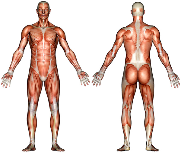
The therapists at Brooklyn Body Works Physical Therapy, P.C. are extremely knowledgeable in all areas regarding health, wellness and fitness.
Head and Neck
TMJ dysfunction
The Temporal Mandibular Joint or TMJ is a joint of the jaw that is made up of two bones (the mandible and the temporal bone of the skull) and a disc that sits between these two bones. The TMJ is essentially the hinge that allows us to open and close our mouths. To find the TMJ, place your fingers on your cheeks just in front of the opening of your ear; open your mouth, you should feel this joint moving. TMJ dysfunction is a usually displacement of this disc, most often in an anterior direction that prohibits normal movement of the joint and may cause pain. In a normally functioning TMJ, the disc should glide anteriorly when you open your mouth, and posteriorly when you close your mouth. The most common symptoms: Crepitus (joint noise, locking, pain and stiffness open when opening or closing the jaw are the 2 most common symptoms. Pain may be felt in the joint, ear, face, jaw, neck or shoulder. Headaches are also a common side effect of this syndrome. Inability to comfortably open the mouth or chew/eat certain foods (for example: biting into a whole bagel, eating an apple) are common problems.
Migraines
A migraine is a very painful headache that has not been proven to have an exact cause. It has been hypothesized to be perhaps due to genetic factors, changes in blood vessels in the brain, or increases in stress levels. The most common symptoms are usually, but not limited to, unilateral (only on one side) head pain, which is often described as “throbbing” and “incapacitating.” The intensity of the headache is moderate to severe and lasts anywhere from 4-72 hours. People with migraines can experience light or sound sensitivity, nausea, or vomiting.
Cervical Strain
A cervical strain is an injury in which the muscles or ligaments of the cervical (neck) area have been stretched or possibly torn, if the injury was severe. It can be due to a motor vehicle accident or just repetitive stress from movement. Most often, a cervical (neck) strain is the result of physical/emotional stress or poor posture.
The most common symptoms: Moderate to severe neck pain (with a feeling of tearing inside the neck if trauma is the cause). Tenderness in the neck, muscle spasms, and stiffness with cervical movements.
Cervical Herniated Nucleus Pulposus (HNP or herniated discs)
The nucleus pulposus is the gel-like center of a spinal disc (the cartilage “cushion” that separates each vertebra in the spine). Its job is to distribute pressure in all directions when the spinal segment is under compression. A Herniated Nucleus Pulposus (HNP) can be called a “slipped disc.” It is when part of the nucleus pulposus is pushed out through a weakened part of the intervertebral disc. In many cases, the nucleus pulposus is expelled in a posterior/lateral (i.e. back and to the side) direction. If this does occur, the herniated nucleus pulposus can push onto the spinal cord or the emerging spinal nerve roots, and cause symptoms depending on the level of herniation. The most common symptoms: Upper extremity weakness, pain or tingling radiating to the shoulder, upper arm, forearm, or perhaps the hand or chest. Most symptoms will only be in one arm, but may occur in both. Pain with rotating or side bending the neck, deep pain over or near the scapula (shoulder blades), headaches.
Facet Joint Syndrome
A facet joint refers to the joints in your spine that connect the vertebra above to the vertebra below to create one complete ‘chain link’ all the way down the spine. The facets are covered with articular cartilage, and have a liquid medium called synovial fluid that lubricates the joint inside the fibrous joint capsule to allow the vertebrae to move without friction. Facet joint syndrome is characterized by pain and inflammation of the facet joints of the spine, usually due to injury (ex: whiplash) or arthritis.
The most common symptoms: Dull, achy pain and tenderness, headaches, neck pain that may radiate into the upper back, pain in the shoulders and limited neck movement (especially rotation).
Shoulder Girdle
Rotator Cuff Injury/Tear
The rotator cuff a group of 4 small, stabilizing muscles that surround and hold the head of your humerus (arm bone) in your shoulder socket. The 4 muscles include: supraspinatus, infraspinatus, subscapularis, and teres minor. Collectively, they provide stability and help with rotational movements of the shoulder joint. A rotator cuff tear is when part of the muscle belly or tendon is torn (can occur in any of these 4 muscles, but most often to the supraspinatus). Commonly, a tear is either due to a specific injury, or “wear and tear” of the muscles from repetitive movements and stresses put on the shoulder joint. Rotator cuff tears occur to people and athletes (ex: baseball players, swimmers) who perform lots of overhead motions or activities.
The most common symptoms: Immediate or gradual onset of pain with movement of the shoulder (depending on mechanism of injury); in time there can be pain even at rest. Pain with lifting the arm overhead or lowering it from a raised position. Weakness with movements, specifically rotation at the shoulder. At times, atrophy (wasting away of the muscle) can be seen.
Adhesive Capsulitis “Frozen Shoulder”
Adhesive Capsulitis “Frozen Shoulder” is a condition that affects middle aged people (often 40-60 years old) characterized by pain and increasing stiffness in the shoulder.
There are three stages that accompany a diagnosis of Adhesive Capsulitis:
1. “freezing”- onset of pain and stiffness
2. “frozen”- limited motion and slow decreases in pain
3. “thawing”- gradual increases in the amount of movement at the shoulder with less and less pain.
The most common symptoms: Dull, achy pain in the shoulder and upper part of the arm. Stiffness and limited or no ability to move the arm, along with incorrect movement patterns.
Shoulder Impingement
An impinged shoulder occurs when there is pressure on the rotator cuff muscles from the acromion (the part of the scapula or “shoulder blade” that sits over the humeral (arm bone) head). With overhead movements of the arm, the acromion pinches or “impinges” upon the rotator cuff muscles. The most common symptoms: The classic symptom is presence of a “painful arc” during lifting the arm. Most often anterior or superior shoulder pain is experienced anywhere from 45° to 120° of elevation.
Elbow / Wrist/ Hand
Lateral Epicondylitis
Often called “tennis elbow” this refers to inflammation from overuse or repetitive stresses on the muscles that attach to the lateral/outside of the elbow joint. There are numerous muscles that extend your wrist and fingers, and all their tendons attach to the outside of the elbow in an area where lots of stress is endured.
The most common symptoms: Tenderness to palpation on the outside of the elbow joint. Pain with functional movements of the forearm, such as grasping or pinching, and specifically resisted wrist extension and wrist radial deviation (bending wrist toward the thumb).
Medial Epicondylitis
Often called “golfer’s elbow” this less common diagnosis refers to inflammation from overuse or repetitive stresses on the muscles that attach to the medial/inside of the elbow joint. This is the opposite of lateral epicondylitis, so the muscles affected are those that help flex the wrist and that move the forearm from a palms up to a palms down position (pronation).
The most common symptoms: Tenderness to palpation on the inside of the elbow joint. Pain with functional movements of the forearm, weak and painful grip strength, and specifically resisted wrist flexion, forearm pronation, and wrist ulnar deviation (bending wrist toward the pinky).
Carpal Tunnel Syndrome:
Carpal Tunnel Syndrome (CTS) is a condition resulting from compression at the wrist of the median nerve (one of the major nerves of the upper extremity). CTS is commonly caused by inflammation (thickening and swelling) of the muscle tendons that also lie in the compartment of the wrist alongside the median nerve.The most common symptoms: Tingling, numbness, or burning sensation in the palm, thumb, and first two fingers. There can be associated decreased grip strength, and symptoms may travel up the arm as well.
Wrist Sprain
A sprained wrist means that ligaments connecting between the bones of the hand and wrist area have become stretched or possibly torn. For example, if you fall backward landing on outstretched palms, you could possibly sprain the ligaments on the palm side of your wrist.
Types: Depending on the severity of the sprain, the doctor may classify you as having a Grade 1, 2, or 3 sprain.
– A grade 1 is mild and the ligaments are only stretched.
– A grade 2 means that some ligaments are badly stretched and others are torn, potentially causing some loss of function in the wrist.
– A grade 3 sprain means that the injury was significant enough to completely tear the ligament from the bone, even pulling a chip of the bone with it (this is called an avulsion fracture).
The most common symptoms: Swelling in the wrist and pain at the time of the injury. Tenderness, bruising, or warmth of the skin over the sprained area. Pain with wrist movements, and a tearing or popping feeling upon injury if the ligaments are torn.
DeQuervain’s Tenosynovitis
Named after a Swiss surgeon, DeQuervain’s Sydrome is a condition classified as inflammation of the tendon sheath (covering) surrounding the two tendons crossing the wrist and connecting to the thumb. As the wrist moves, the numerous tendons of finger and wrist muscles get pulled along and move within their individual compartments within the wrist. Especially with repetitive wrist or thumb movements, there can be irritation of these tendon sheaths, causing them to swell and enlarge, therefore causing pain. The most common symptoms: Pain and tenderness on the thumb side of the wrist in the area where the thumb bone and radial bone of the forearm meet. A “sticking” or locking and creaking of the thumb with movement. Pain that increases with forceful grasping, lifting of heavy objects, making a fist, or pinching motions. Flexing the thumb into the palm, folding the 4 fingers over it into a fist, and bending the wrist toward the pinky causes pain for people who have this condition.
Mid and Lower Back
Disc Herniation
A disc refers to the cartilage “cushion” that separates each vertebral bone in the spine. The disc is gel-like and acts as a shock absorber to distribute forces throughout the body. A disc herniation means that part of the intervertebral disc has been forced outside of the spinal column. Usually the herniation is due to some type of trauma (often lifting) and the disc is pushed posteriorly. It compresses the spinal nerve roots that are exiting the spinal cord at a particular level. The most common symptoms: Pain felt in the back, often traveling down the legs on one or both sides of the body. There can be sensory or motor changes too.
Muscle Spasm and Strain
A muscle spasm is an involuntary (not under your control) contraction of a muscle. These spasms can last from seconds to hours. A spasm is the resulting inflammation that occurs after a muscle strain. A muscle strain, often referred to as a “pulled muscle,” this means that either the muscle belly or the tendon (the part of the muscle that attaches to bone) is injured. It can be overstretched or torn. The most common symptoms: Inflammation (swelling, redness) in the area of the muscle. Tenderness or weakness when using or attempting to use the muscle. Pain at rest and when using the muscle.
Arthritis or Osteoarthritis
Arthritis refers to the wearing and tearing away of the cartilage (tissue covering the ends of bones) in joints of the body that occurs as a result of the normal aging process, injury to the joint, or misuse. As the cartilage wears down, the bones rub against each other with movement, and since they don’t have the protection and shock absorption from the now worn down cartilage, the joint can become damaged. Arthritis is typically seen in the joints of the hands, knees, hips, and vertebrae of the spine.
Osteoarthritis, a type of arthritis, refers specifically to bony changes from wear and tear. Overtime there is loss of cartilage in the joint, overgrowth of bone, and development of bony spurs (abnormal bony projections in the joint space making movement more difficult and painful).
The most common symptoms: Stiffness, pain, swelling in the joint, and lack of full range of motion.
Hip/ Pelvis
Labral Tear
The labrum is cartilage of the hip joint, where the head of the femur (large thigh bone) sits in the socket of the pelvis, called the acetabulum. The labrum lines this socket to provide cushion and to help stabilize the femur in place during movement.
A tear of this labral cartilage results from injury, repetitive motions causing wear and tear on the hip joint, or from degenerative processes like osteoarthritis. The most common symptoms: A locking, catching or a painful clicking in the hip with movements. Limited range of motion and stiffness can result. Other times, there may be no symptoms associated with a labral tear.
Bursitis of the hip (a.k.a. Trochanteric bursitis)
As mentioned earlier, a bursa is a small fluid-filled sac that cushions a number of joints in the body to decrease pressure points between bone and muscle tendons to allow smooth movements. With overuse, repetitive movements, or injury of falling onto that hip, the bursa between the ITB and greater trochanter of the femur can get inflamed. With inflammation, the bursa can enlarge and can cause pain every time the tendon moves over the bone. In the hip joint, bursitis is common in athletes. The most common symptoms: Sharp pain with hip movements, specifically flexing and extending. Tenderness to touch over the area, and swelling. There is often more pain at night, especially lying on the affected hip, or pain with prolonged walking or stair climbing.
Knee and Thigh
ITB Syndrome
ITB syndrome is an overuse injury that occurs as a result of repetitive rubbing of the ITB over the lateral condyle of the femur. However, as the knee is repetitively flexed and extended, such as with running, friction is often created over this bursa causing irritation and inflammation. It commonly occurs in long distance runners, walkers and cyclists. Predisposing factors include pronated feet, “bowed” or “knock” knees and muscle imbalances of the hip and thigh. It is very common to occur in long distance runners, walkers, bicyclists, or due anatomical abnormalitites like overly pronated feet or bow-legs, or muscle imbalances like a weak gluteus medius muscle of the buttocks. The most common symptoms: Pain on the outside of the knee just above the joint during and after activity, and increased thickening of the tissue and tenderness. There may be a snapping sensation felt on the outside of the knee and pain may travel up or down the leg.
Sciatica
Ultimately sciatica refers to a group of symptoms that present as low back or buttock pain, with pain traveling down the back of the leg. It occurs when there is compression either of one of the nerve roots that make up the sciatic nerve or compression of the sciatic nerve itself. The sciatic nerve is the longest and widest nerve in the body, and it is made from spinal nerve roots L4-S3. Because this nerve courses below (or on rare occasions through) the piriformis muscle in the buttock, tightness of the piriformis muscle can cause sciatica. Other causes include spinal disc herniation, spinal stenosis (stiffening of the spine), or degenerative disease. The most common symptoms: Pain from the lower back or buttock region that travels down the back of one leg of varying severity. There can also be tingling, numbness, or progressive weakness in the leg.
ACL injury
The Anterior Cruciate Ligament (ACL) is one of the four major ligaments that provide stability to the knee. It connects the posterior part of the femur (large thigh bone) to the anterior part of the tibia (“shin bone”) and resists anterior translation of the tibia on the femur. Essentially, it is responsible for keeping the two bones in alignment so the tibia does not move excessively forward while the femur is not moving. An ACL tear is when the ligament is torn (often during a sporting incident) from increased torsion at the knee joint, a sudden dislocation or hyperextension of the knee. In many cases, the ACL is torn when the foot is planted, the knee is locked, and there is a rapid change of direction putting increased forces on the knee.
The most common symptoms: A “popping” sound heard or felt initially upon injury, followed by pain at the outside and back of the knee, swelling, and a feeling of instability around the knee with movement. Also knee flexion and extension may be painful and limited.
Meniscal injury
A meniscus is one of two strong, fibrous, “C” shaped cartilage discs that sit between the femur and the tibia in the knee. The medial and lateral menisci act as shock absorbers and help lubricate the joint slightly, therefore acting as a buffer between the two bones. A meniscal tear occurs from one of two reasons, injury from trauma (like in sports) from the leg twisting on a bent knee, or from degeneration in old age. The most common symptoms: A “pop” heard upon traumatic injury, a clicking sound with knee movements, inability to completely straighten the leg because the knee feels “locked.” Also there is pain and swelling in the knee joint, and tenderness to palpation of the joint line between the femur and the tibia, which is where the menisci are located.
Patellofemoral Syndrome
Patellofemoral refers to the knee where the patella (kneecap) and femur (thigh bone) approximate. Repeated flexion and extension of the knee can cause irritation as these two bones are constantly rubbing against each other to allow movement. Patellofemoral Pain Syndrome is characterized by pain behind and/or around the kneecap. It is also generally known as “anterior knee pain” (AKP). It is likely due to physical or biomechanical changes that occur in this joint as a result of overuse, repetitive motions, muscular imbalances or weight increases. Ultimately, the greater the contact between the patella and the femur overtime, the greater someone’s chance is of developing this syndrome. The most common symptoms: Anterior knee pain is the most common complaint, a pain felt right behind the knee cap. Also the pain seems to get worse with activity and going down stairs. There can be a grinding sensation felt behind the patella, and it often gets worse with prolonged sitting.
Lower Leg/ ankle and foot
Achilles Tendonitis
The Achilles tendon is the thickest and strongest tendon in the body that serves as the attachment of the calf muscles (gastrocnemius and soleus muscles) to the heel bone on the backside of the lower leg behind the ankle. Its job is to assist with walking and lifting the heels off the ground. Achilles Tendonitis is inflammation of the Achilles tendon. This condition is common in women who wear high heels daily, since this causes the calf muscles and the Achilles tendon to develop a shortened position. When the woman wears flat shoes, the increased stretch on these structures causes pain. The most common symptoms: Gradual pain in the lower calf above the heel, that worsens with prolonged walking or continual activity. Pain could occur when wearing flat shoes after many hours of high heels.
Plantar Fasciitis
The plantar fascia helps maintain the arch of the foot. Plantar Fasciitis is a painful, inflammatory condition where the plantar fascia becomes inflamed due to excessive stresses put on the fascia, or biomechanical problems causing increased pronation of the foot. Poor arch support, flat shoes, inactivity, long periods of weight bearing, or weight gain can all cause plantar fasciitis to occur. The most common symptoms: Intense pain at the bottom of the foot, with the most tender spot being at the base of the heel toward the arch, The pain is most severe upon waking up in the morning and the initial first steps after a period of prolonged sitting.
Sprained Ankle (Inversion Ankle Sprain)
A sprained ankle commonly occurs following a sideways movement of the foot, like landing incorrectly from a jump or on uneven terrain. The majority of ankle sprains are caused by inversion motion of the ankle (when the bottom of the foot turns inward toward the opposite leg). The outer ligaments of the ankle become overly stretched and possibly torn. The most common symptoms: Pain on the outer part of the ankle (with an inversion sprain) and swelling. Bruising, ankle instability, and difficulty walking is common.
Morton’s Neuroma
Morton’s Neuroma is a thickening or enlargement of the tissue surrounding one of the nerves under the foot as it is formed from two smaller nerves (the medial and lateral plantar nerves) between the 3rd and 4th toes. Since the nerve(s) sits just above the fat pads in the bottom of your feet, they may become compressed with each step. From above, it gets pushed down on by a strong ligament responsible for holding together the bones of the foot (the deep transverse metatarsal ligament) and from below, it gets pushed up on by the ground. Irritation of the nerve is commonly due to excessive pressure in the toe region or trauma. The most common symptoms: Localized pain between the 3rd and 4th toes and often at the ball of the foot. Pain becomes more severe with weight bearing and wearing shoes (especially high heels or shoes with narrow toe boxes). Pain is usually relieved with barefoot or at night.
Bunions
A bunion is a bump on the outside of your big toe joint that is either due to a structural bony deformity or inflammation of the tissues at the joint. As a result, the big toe turns and leans in toward the other toes of the foot, thus creating an angle between the inside line of the foot (along the arch) and the tip of the big toe. This deformity can be genetic or due to poor biomechanics of the foot, such as flat feet and poorly fitting or tight shoes. The most common symptoms: A distinct bump on the outside of the big toe that is possibly swollen, red, tender, and painful. There is pain with walking, as the big toe joint extends with every step. Depending on how long you’ve had a bunion the big toe may be only slightly or very turned inward toward the other toes.
Hammer Toe
Hammer toes are deformities in the toes (usually toes 2-5) in which there is a muscle/tendon imbalance. As a result, the muscle/tendons of the foot lose mechanical advantage to work in the way they normally would. The classic sign of a hammer toe deformity is where the middle toe joint points up and the farthest tip of the toe points down, almost creating an upside-down “V” of your toe. This can result from injury to one of the tendons, having a second toe longer than the big toe, or from long periods of wearing narrow shoes that cramp the toes or high heels that hold the toes in one position with lots of pressure for a long time. After a while, the tendons on top of the foot cannot straighten the toe out. The most common symptoms: A callus or corn that has formed on the top of the middle toe joint, pain over the top of the toe, and inability to actively, fully straighten the toe.


